Rib Cage Anatomy Diagram / Two main divisions of the human skeleton - Online Science ... - Go back to the diaphragm area and use a scalpel to cut the wall of the body cavity away from the diaphragm.
Rib Cage Anatomy Diagram / Two main divisions of the human skeleton - Online Science ... - Go back to the diaphragm area and use a scalpel to cut the wall of the body cavity away from the diaphragm.. The human lungs are found in the chest, within the thoracic cavity. The thoracic cavity contains the lungs and the heart, which is located in the mediastinum. The urinary system is located directly below the rib cage. Human skeleton, the internal skeleton that serves as a framework for the body. Urinary system diagram what is a urinary system diagram?
Gross anatomy normally, the aorta ascends in the superior medias. The thoracic cavity is the more superior subdivision of the anterior cavity, and it is enclosed by the rib cage. This framework consists of many individual bones and cartilages. Several muscles that move the arms, head, and neck have their origins on the sternum. Urinary system diagrams are illustrations of the urinary system, also referred to as the renal system.
Front view of rib cage and spine on black background ... from st.focusedcollection.com Urinary system diagrams are illustrations of the urinary system, also referred to as the renal system. An inhalation is accomplished when the muscular diaphragm, at the floor of the thoracic cavity, contracts and flattens, while the contraction of intercostal muscles lift the rib cage up and out. This framework consists of many individual bones and cartilages. Variant anatomy of the aortic arch occurs when there is failure of normal aortic development. Jan 01, 2019 · the kidneys and lungs are located in the back, with the lungs found within the rear portion of the rib cage and the kidneys situated near muscles just below the cage. Human skeleton, the internal skeleton that serves as a framework for the body. The external intercostals elevate the ribs during forced inspiration, expanding the thorax and lungs. It results in a number of heterogenous anomalies of the aorta and its branch vessels.
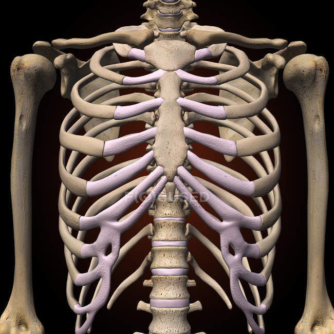
Jul 16, 2019 · the sternum, commonly known as the breastbone, is a long, narrow flat bone that serves as the keystone of the rib cage and stabilizes the thoracic skeleton.
The urinary system is located directly below the rib cage. Variant anatomy of the aortic arch occurs when there is failure of normal aortic development. It results in a number of heterogenous anomalies of the aorta and its branch vessels. The spinal cord is about 18 inches (45 centimeters) in length and is relatively cylindrical in shape. Jul 16, 2019 · the sternum, commonly known as the breastbone, is a long, narrow flat bone that serves as the keystone of the rib cage and stabilizes the thoracic skeleton. The diaphragm should remain intact, but now the rib cage can be pulled back and pinned to the pan, exposing the thoracic cavity. However, they also have additional individual functions. There also are bands of fibrous connective tissue—the ligaments and the tendons—in intimate relationship with the parts of the skeleton. The human rib cage is a component of the human respiratory system. Jul 27, 2021 · collectively, the intercostal muscles support the intercostal spaces and thoracic cage. An inhalation is accomplished when the muscular diaphragm, at the floor of the thoracic cavity, contracts and flattens, while the contraction of intercostal muscles lift the rib cage up and out. Anatomically, the spinal cord runs from the top of the highest neck bone (the c1 vertebra) to approximately the level of the l1 vertebra, which is the highest bone of the lower back and is found just below the rib cage. Go back to the diaphragm area and use a scalpel to cut the wall of the body cavity away from the diaphragm.
Go back to the diaphragm area and use a scalpel to cut the wall of the body cavity away from the diaphragm. Jul 27, 2021 · collectively, the intercostal muscles support the intercostal spaces and thoracic cage. The thoracic cavity contains the lungs and the heart, which is located in the mediastinum. When you reach the midpoint between the forelegs, make another incision down to the pan. Human skeleton, the internal skeleton that serves as a framework for the body.
Rib cage - skeletal system | Lpn to rn programs, Medical ... from i.pinimg.com Variant anatomy of the aortic arch occurs when there is failure of normal aortic development. In contrast, the internal and innermost intercostals depress the rib cage during forced expiration. The thoracic cavity is the more superior subdivision of the anterior cavity, and it is enclosed by the rib cage. When you reach the midpoint between the forelegs, make another incision down to the pan. There also are bands of fibrous connective tissue—the ligaments and the tendons—in intimate relationship with the parts of the skeleton. Anatomically, the spinal cord runs from the top of the highest neck bone (the c1 vertebra) to approximately the level of the l1 vertebra, which is the highest bone of the lower back and is found just below the rib cage. It results in a number of heterogenous anomalies of the aorta and its branch vessels. An inhalation is accomplished when the muscular diaphragm, at the floor of the thoracic cavity, contracts and flattens, while the contraction of intercostal muscles lift the rib cage up and out.
Jan 01, 2019 · the kidneys and lungs are located in the back, with the lungs found within the rear portion of the rib cage and the kidneys situated near muscles just below the cage.
In contrast, the internal and innermost intercostals depress the rib cage during forced expiration. Urinary system diagrams are illustrations of the urinary system, also referred to as the renal system. This framework consists of many individual bones and cartilages. It encloses the thoracic cavity, which contains the lungs. The thoracic cavity is the more superior subdivision of the anterior cavity, and it is enclosed by the rib cage. The urinary system, at a high level, contains two kidneys, two ureters, a urethra, and a bladder. The urinary system is located directly below the rib cage. Jul 16, 2019 · the sternum, commonly known as the breastbone, is a long, narrow flat bone that serves as the keystone of the rib cage and stabilizes the thoracic skeleton. The human lungs are found in the chest, within the thoracic cavity. The left lung is somewhat smaller than the right lung. When you reach the midpoint between the forelegs, make another incision down to the pan. It results in a number of heterogenous anomalies of the aorta and its branch vessels. The diaphragm should remain intact, but now the rib cage can be pulled back and pinned to the pan, exposing the thoracic cavity.
Gross anatomy normally, the aorta ascends in the superior medias. The external intercostals elevate the ribs during forced inspiration, expanding the thorax and lungs. Urinary system diagrams are illustrations of the urinary system, also referred to as the renal system. Anatomically, the spinal cord runs from the top of the highest neck bone (the c1 vertebra) to approximately the level of the l1 vertebra, which is the highest bone of the lower back and is found just below the rib cage. Go back to the diaphragm area and use a scalpel to cut the wall of the body cavity away from the diaphragm.
Vintage Anatomy Print Features The Side View Of The Human ... from media.gettyimages.com Go back to the diaphragm area and use a scalpel to cut the wall of the body cavity away from the diaphragm. Variant anatomy of the aortic arch occurs when there is failure of normal aortic development. The urinary system, at a high level, contains two kidneys, two ureters, a urethra, and a bladder. The thoracic cavity is the more superior subdivision of the anterior cavity, and it is enclosed by the rib cage. There also are bands of fibrous connective tissue—the ligaments and the tendons—in intimate relationship with the parts of the skeleton. Jul 16, 2019 · the sternum, commonly known as the breastbone, is a long, narrow flat bone that serves as the keystone of the rib cage and stabilizes the thoracic skeleton. Anatomically, the spinal cord runs from the top of the highest neck bone (the c1 vertebra) to approximately the level of the l1 vertebra, which is the highest bone of the lower back and is found just below the rib cage. When you reach the midpoint between the forelegs, make another incision down to the pan.
The urinary system is located directly below the rib cage.
Urinary system diagram what is a urinary system diagram? The diaphragm should remain intact, but now the rib cage can be pulled back and pinned to the pan, exposing the thoracic cavity. Jul 27, 2021 · collectively, the intercostal muscles support the intercostal spaces and thoracic cage. The urinary system, at a high level, contains two kidneys, two ureters, a urethra, and a bladder. Variant anatomy of the aortic arch occurs when there is failure of normal aortic development. However, they also have additional individual functions. Go back to the diaphragm area and use a scalpel to cut the wall of the body cavity away from the diaphragm. It encloses the thoracic cavity, which contains the lungs. The thoracic cavity is the more superior subdivision of the anterior cavity, and it is enclosed by the rib cage. The left lung is somewhat smaller than the right lung. Jul 16, 2019 · the sternum, commonly known as the breastbone, is a long, narrow flat bone that serves as the keystone of the rib cage and stabilizes the thoracic skeleton. The human lungs are found in the chest, within the thoracic cavity. The external intercostals elevate the ribs during forced inspiration, expanding the thorax and lungs.
Anatomically, the spinal cord runs from the top of the highest neck bone (the c1 vertebra) to approximately the level of the l1 vertebra, which is the highest bone of the lower back and is found just below the rib cage rib cage anatomy. When you reach the midpoint between the forelegs, make another incision down to the pan.


0 Komentar